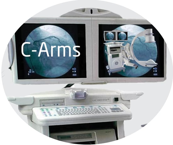
In the past few years, C-Arm technology has dramatically improved. Mobile C-Arms are delivering the  same high quality images that costly fixed-arm platforms are delivering and as a whole, they’re getting easier and easier to use. Let’s take a closer look at what’s new in C-Arm technology.
Imaging is awesome. Flat panel CMOS image detectors are allowing surgeons to magnify images without increasing radiation dose or losing clarity. By using low-dose radiation, they are capturing superior high quality images. Want to zero in on small anatomical landmarks and display captured images on large 4K monitors? It’s easy with today’s newer systems.
New cooling systems make a big difference. It’s no secret that C-Arms are the workhorse of medical imaging devices. However, when used for hours on end overheating can be an issue, When C-Arms go offline because of heating, procedures grind to a halt until a tech can retrieve a replacement machine or the unit cools off, especially during intense orthopedic or spine cases. However, C-Arms with advanced liquid cooling systems maintain a constant circulation of fluid through an imaging tube and cooling reservoir, which prevents overheating and keeps the system working.
Easy, peasy. The newest C-Arms have smaller footprints, so the imaging units take up less space in outpatient ORs. In addition, improved motorized mechanisms deliver pinpoint control when placing the C-Arm into position. There are also smart anti-collision features that sound the alert if the arm is close to coming in contact with the table or even a patient. It will also shut down if it’s moved any closer.
Newer C-Arms also have user-friendly touchscreen controls, so programming settings between cases is intuitive and quick. Anatomic- and procedure-specific profiles allow you to calibrate C-Arms for cases with the touch of a button. In addition, the profiles change the scale of the captured images and adjust contrast in order to optimize the unit for pediatric, abdominal or extremity surgeries. The settings automatically adjust the radiation dose as well, differentiating between the amount need in order to capture images during major spine procedures and extremity surgeries. There are also “position save and recall” features that allow surgeons to set an arm to an exact position, save the position and return to it within mere millimeters —with just the push of a button.
Managing your images. This is much easier, too, thanks to advanced software systems on newer machines that capture digital images for storing or sending to picture archiving and communication systems through a secure network or for storage.
Viewing in 3D. While 3D imaging of targeted anatomy hasn’t been clinically proven to improve overall surgical outcomes, it can help surgeons better reduce distal radial joint fractures. The field of view delivered by 3D C-Arms lets surgeons achieve the axial cut. This helps them reduce a tibia or make sure the reduction is properly placed during repairs of tibial plateau fractures.
Talk To An Expert
Yes, new C-Arm technology is exciting. However, if you’re looking for new medical imaging equipment, oftentimes you can get the performance you want with a refurbished or used system—at a fraction of the cost. Atlantis Worldwide has been helping clients find ideal imaging solutions for more than 29 years. Talk with one of our experts today.
Some blogs you may have missed:
- Mini C-Arms Costs & Comparisons
- C-Arms & Vascular Health
- Philips C-Arm Manufacturer Spotlight
- Top 7 C-Arms for Your Orthopedic Practice
- C-Arm Articles
About the author: Vikki Harmonay


