Case Study: Role of image-based iterative reconstruction technique for low radiation dose chest CT examination
Sarvenaz Pourjabbar, MD; Ranish Deedar Ali Khawaja, MD; Sarabjeet Singh,MD; Atul Padole, MD; Diego Lira, MD; Mannudeep Kalra, MD
Summary
A 50-year-old male weighing 80 kg (body mass index: 22 kg/m2) underwent follow up chest CT for further evaluation of indeterminate focal ground glass opacity in the right lower lobe. The patient had symptoms of shortness of breath, fatigue, chest pain and pressure with exertion that were unrelated to coronary artery disease. In addition, the patient had history of progressive muscular weakness and difficulty in walking, for which a skeletal muscle biopsy was performed and revealed chronic neuropathic myopathy. The patient was suspected to have paraneoplastic syndrome and for which he underwent the initial chest and abdominal CT.
Diagnosis
Pneumonia or aspiration. Given resolution of the focal opacity in the right lower lobe, the finding likely represented a small focus of pneumonia or aspiration.
 Findings
At the time of follow up chest CT, written informed consent was obtained from the patient for acquiring a low dose CT image series in addition to the standard of care chest CT. Standard of care chest CT was acquired on a 64-section multidetector-row CT scanner (Discovery 750HD, GE Healthcare) at 400 mA. Immediately after the standard, we acquired low dose CT images over a 10 cm scan length in the mid chest and reduced tube current of 80 mA. Other scan parameters were held identical to the standard of care CT at 120 kV, pitch of 0.984:1, table speed 40 mm per rotation, 64*0.625 mm detector configuration, and 0.5 second gantry rotation time. All standard of care and research images were reconstructed at 2.5 mm section thickness and 2.5 mm section increment using detail reconstruction algorithm using conventional filtered back projection technique. The volume CT dose index (CTDIvol) for standard of care and research low dose CT series was 15 mGy and 3.4 mGy, respectively, resulting in 75% radiation dose reduction. The research images were then processed with SafeCT algorithm in order to improve the image quality.
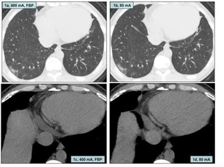
SafeCT processed low dose CT images (Figure 1) at 75% lower radiation dose were then compared with the standard of care CT images. Both the processed low dose and unprocessed standard of care CT images demonstrated patchy ground glass opacity in the superior segment of the right lower lobe. No significant mediastinal or hilar lymphadenopathy was noted on either set of images. Diagnostic confidence and image quality on both sets of images were identical. Subsequent follow up chest CT showed interval resolution of the ground glass opacity in the right lower lobe.
Discussion
Several studies have evaluated strategies to reduce radiation dose from CT such with reduction of tube potential and tube current, and with improvement in image quality of low radiation dose images with image noise reduction filters and iterative reconstruction techniques [1-4]. Iterative reconstruction algorithms reduce image noise while maintaining or improving other image attributes (such as sharpness and contrast) in the low dose CT images [5-9]. The Food and Drug Administration (FDA) approved three-dimensional (3D) image-based iterative reconstruction algorithm, SafeCT used in our study segments CT images into 3D patches to estimate the noise statistics and signal. Then, images are processed in an iterative loop to reach an acceptable quality [10]. Since this process takes place entirely in DICOM image domain, it can process images from any CT vendor and takes few seconds to process a routine CT examination. Generally, SafeCT image processing server lies between the CT scanner and the PACS so that images are processed with SafeCT when they arrive to the interpretation workstations in an automatic manner [10].
The example presented in this article demonstrates significant potential of image based iterative reconstruction techniques such as SafeCT for dose reduction while retaining the image quality and diagnostic information.
Conclusion
Radiation dose reduction for chest CT is important and feasible with use of iterative reconstruction techniques, which help improve image quality of low dose CT images.
Figure legend
Figure 1: Follow up chest CT examination was performed at standard (400 mA, fig 1a and 1c) and low dose (80 mA, fig 1b and 1d) and the low dose images were processed with image based iterative reconstruction technique. Lung window shows ground glass opacity in right lower lobe in transverse chest CT images at both dose levels as well as the pericardium in the mediastinal window settings.
References
1) Kalra MK, Maher MM, Toth TL, Hamberg LM, Blake MA, Shepard JA, Saini S. Strategies for CT radiation dose optimization. Radiology. 2004 Mar;230(3):619-28.
2) Singh S, Kalra MK, Moore MA, Shailam R, Liu B, Toth TL, Grant E, Westra SJ. Dose Reduction and Compliance with Pediatric CT Protocols Adapted to Patient Size, Clinical Indication, and Number of Prior Studies. Radiology. 2009 Jul;252(1):200-8.
3) Kalra MK, Singh S, Thrall JH, Mahesh M. Pointers for optimizing radiation dose in abdominal CT protocols. J Am Coll Radiol. 2011 Oct;8(10):731-4.
4) Singh S, Kalra MK, Thrall JH, Mahesh M. Pointers for optimizing radiation dose in chest CT protocols. J Am Coll Radiol. 2011 Sep;8(9):663-5.
5) Singh S, Kalra MK, Gilman MD, Hsieh J, Pien HH, Digumarthy SR, Shepard JA. Adaptive statistical iterative reconstruction technique for radiation dose reduction in chest CT: a pilot study. Radiology. 2011 May;259(2):565-73.
6) Singh S, Kalra MK, Hsieh J, Licato PE, Do S, Pien HH, Blake MA. Abdominal CT: comparison of adaptive statistical iterative and filtered back projection reconstruction techniques. Radiology. 2010 Nov;257(2):373-83.
7) Singh S, Kalra MK, Shenoy-Bhangle AS, Saini A, Gervais DA, Westra SJ, Thrall JH. Radiation Dose Reduction with Hybrid Iterative Reconstruction for Pediatric CT. Radiology. 2012 May;263(2):537-46.
8) Singh S, Kalra MK, Do S, Thibault JB, Pien H, Connor OO, Blake MA. Comparison of hybrid and pure iterative reconstruction techniques with conventional filtered back projection: dose reduction potential in the abdomen. J Comput Assist Tomogr. 2012 May;36(3):347-53. PubMed PMID: 22592622.
9) Kalra MK, Woisetschläger M, Dahlström N, Singh S, Lindblom M, Choy G, Quick P, Schmidt B, Sedlmair M, Blake MA, Persson A. Radiation dose reduction with sinogram affirmed iterative reconstruction technique for abdominal computed tomography. J Comput Assist Tomogr. 2012 May;36(3):339-46. PubMed PMID: 22592621.
10) Pourjabbar S, Singh S, Singh AK, Johnston JP, Shenoy-Bhangle A,Do S, Padole A, Blake MA, Persson A, Kalra MK. Prospective clinical study to assess image based iterative reconstruction for abdominal CT acquired at two radiation dose levels. Accepted JCAT June 3rd 2013.
Follow Atlantis Worldwide on Twitter: @AtlantisLLC
Other blogs you may have missed:
- Is Your CT Tube About To Fail?
- Should your business lease or buy medical imaging equipment?
- Extend The Life of Your Medical Imagining Equipment or Replace It?
- Plan Ahead For Medical Imaging Equipment Purchases


