The world of medical imaging devices is ever changing. Case in point: more and more portable ultrasound machines 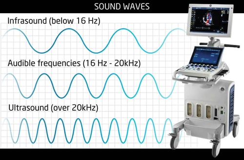 are being used in a wide variety of applications—and for good reasons. They’re extremely easy to use, portable, lightweight, compact and deliver good quality images. Their portability makes them very convenient, and their batteries deliver long life both on and off site.
Portable ultrasound machines are available in a wide range of frequencies, from 3-20 MHz, making them suitable for a wide range of applications, from Abdominal, Nephrology, Orthopedics, Podiatry and Rheumatology to Gastroenterology, Cardiology, Urology, Vascular/Peripheral vascular and more.
Let’s take a look at the variances of ultrasound frequencies in portable radiology equipment.
Sound waves are a type of mechanical energy that can be used to produce images of our bodies’ internal structure and organs through ultrasound technology. However, sound can’t travel without a medium and the velocity of sounds waves don’t remain constant. A sound beam becomes narrower and more directional with an increase in its frequency. It’s also important to note than the frequency of sound waves have to be inversely proportional to their penetration. The penetration of a sound wave decreases with an increase in frequency, and vice versa.
When sound waves pass through a medium, it causes vibrations in their molecules. When the molecules expand and contract, the motion of sound waves pass from one molecule to the next.
The velocity of a sound wave isn’t constant. It changes with the compressibility of the medium through which it travels and is directly correlated with the firmness of the medium. Gases have the highest compressibility, solids have the lowest and liquids fall in between. When a sound wave passes from a liquid into a gas or from a gas to a liquid, its velocity increases, regardless of the frequency.
In order to help medical imaging professionals select the right ultrasound frequency for imaging a body part, they need to keep this in mind: High-frequency sound waves are absorbed easily and have shorter wavelengths than low frequency sound waves, so they can’t penetrate as deep as low-frequency waves. As a result, higher ultrasound frequencies are typically used to image superficial body structures and low ultrasound frequencies are used to scan deeper organs and structures.
All portable ultrasound machines come with a probe or scanner that is to be placed on area of patient to be examined. A gel is applied to area for easy transmission of sound waves. A diagnostic ultrasound transducer can operate at more than one frequency.
Here is a guide to the frequencies and the body parts:
2.5 MHz: obstetric, gynecological and deep abdomen imaging
3.5 MHz: obstetric, general abdomen and gynecological imaging
5 MHz: vascular, breast and general abdomen imaging
7.5 MHz: breast and thyroid
10 MHz: thyroid, breast, superficial masses and veins and musculoskeletal imaging
15 MHz: musculoskeletal and superficial structures imaging
Talk To An Expert
If your practice, hospital, clinic or healthcare facility is thinking of purchasing a new portable diagnostic imaging system, it’s wise to talk to an expert, like Atlantis Worldwide. Oftentimes refurbished or used portable diagnostic radiology equipment can be ideal and offer lower prices and impressive warranties. Contact one of our experts today!
Some blogs you may have missed:
- Plan Ahead For Medical Imaging Equipment Purchases
- Should your business lease or buy medical imaging equipment?
- Changing the Game: Xbox to X-Ray
- 7 Benefits of Medical Imaging File Sharing
- 3D Printing in Medical Imaging & Healthcare
- X-Ray - Nuclear - PET/CT Articles
Meet the author: Vikki Harmonay