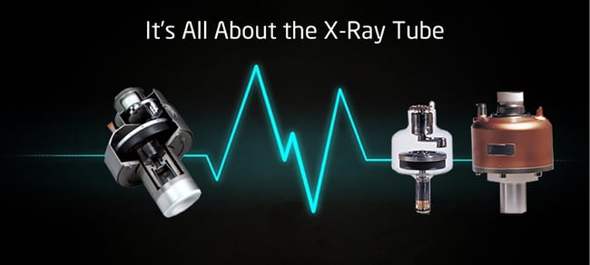
X-ray tubes produce x-ray photons that then create the CT image. Their design is a modification of a standard rotating anode tube  like those used in angiography. Tungsten, which has an atomic number of 74, is usually used for the anode target material, which produces a higher intensity x-ray beam because the intensity of x-ray production is approximately proportional to the atomic number of the target material.
CT scanner tubes often contain multiple sizes of focal spots—usually 0.5 and 1.0. Small focal spots in Computed Tomography tubes produce sharper images because of reduced penumbra, like in standard x-ray tubes. They concentrate heat onto smaller areas of the anode that can’t tolerate as much of the heat.
Scanning protocols often require multiple long exposures performed on numerous patients per day which creates a lot of stress on the tube. Manufacturers list generator and tube cooling capabilities in their product specifications. The generator’s maximum power is usually listed in kW while the anode heat capacity is listed in million heat units MHU and the maximum anode heat dissipation rate is listed in thousand heat units KHUY. These specifications can help differentiate various CT Scanner systems. Remember, these values represent the highest limit of tube performance. There are also guidelines to the length of protocols that the tube will allow, as well as how quickly those protocols can be repeated.
About Filtration
In order to shape the x-ray beam, compensating filters are used. These reduce the radiation dose to the patient and help minimize image artifact. Radiation emitted by a CT x-ray tube is polychromatic. By filtering the x-ray beam, the range of x-ray energies that reach the patients are reduced. The filtering removes the long wavelength or “soft rays,” which are readily absorbed by the patient and don’t contribute to the CT image. When you create a more uniform beam intensity by reducing artifacts that result from beam hardening, it improves the CT image.
Different filters are used when scanning the body versus the head. When scanning the human anatomy, you get a thicker round cross section in the middle than along the edges. As a result, body scanning filters are used to reduce the beam intensity at the periphery of the beam, corresponding to the thinner areas of a patient’s anatomy. These are often referred to as “bow tie areas” because of their shape.
About Collimation
Collimation restricts the x-ray beam to a specific area, which helps reduce scatter radiation. It controls the slice thickness by narrowing or widening the x-ray beam. It’s located near the X-Ray Machine.
Scatter radiation can reduce image quality and increase the patient’s radiation dose. By reducing the scatter radiation, you can get better contrast resolution and decrease patient dose, or the amount of x-ray beam before it passes through the patient.
This is Part Two of CT Components.
In part three you will learn everything about Detectors. In case you missed part one-this is what it contained: The Gantry, Slip Rings, Cooling System and the Generator- click here to read it!
Talk To An Expert
If you have questions about Refurbished CT Scanner systems or their components, talk to an expert at Atlantis Worldwide. We’ve been providing used and refurbished medical imaging equipment to hospitals, clinics, urgent care facilities, veterinary clinics and other healthcare practices for more than 27 years and would be happy to help you.
Some blogs you may have missed:
- Should You Buy New Or Used X-Ray and CT Tubes?
- CT Scanner Artifacts- How Do I Correct Streaks?
- The Lowdown on CT & X-Ray Replacement Tubes
- Is Your CT Tube About To Fail?
- Free CT Resources
About the author: Vikki Harmonay


