You never know when inspiration will strike, as proven in the 60s, when Research Engineer Godfrey Hounsfield had a conversation with a doctor while on vacation.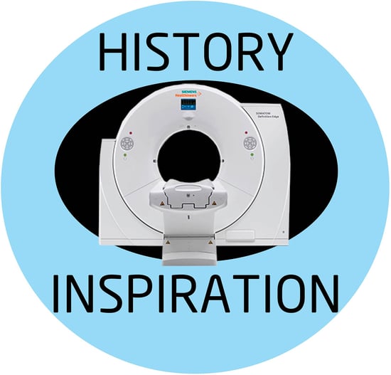 The physician shared his frustration about the quality of X-ray images of the brain being too grainy and two-dimensional.
It just so happened that Hounsfield worked for Electrical and Musical Industry in Hayes, England, which developed electronic equipment, but was best known for producing and selling records by the Beatles.
Post vacation, the engineer approached his supervisor about trying to develop a machine that could create three-dimensional images of the brain. He believed he could create a machine that would send narrow beams of X-rays through an individual’s head, with a computer using the resulting data to construct a series of cross sections that when assembled together, would create a 3D image of the brain.
He worked with a team of neurologists to build the machine. In 1971, the first computed tomography scan of the human brain was created. As a result, Hounsfield was a co-recipient of the Nobel Prize in 1979 in Physiology or Medicine.
Hounsfield’s journey to the CT Scanner was an interesting one. He first served with the Royal Air Force, learning the basics of radar and electronics. He joined EMI in 1951, developing radar and guided weapons systems. In 1958, he helped design Britain’s first commercially available all-transistor computer called the Emidec 1100. His boss warned him that his job would be in jeopardy if he didn’t develop another great idea—which ended up being the CT Scanner. EMI didn’t want to get into the medical imaging business but was willing for Hounsfield to work on the CT Scanner, as long as he could raise additional funding. He applied for and received a $40,000 grant from the British Department of Health and Social Care.
The first prototype was small enough to fit on a table top, and was tested on small pigs. Once the scanner was successful, a full-sized scanner was built to test on human brains that had been preserved in formalin. Unfortunately, the formalin hardened the brain tissue so it didn’t even look like normal brain matter when scanned. The scientists then tried cow brains, but since they had been killed with an electric shock, the brains were filled with blood, obstructing the radiologists’ view of the organ’s structure and mass.
A team member was Jewish and suggest using kosher cow brains since they died by having their jugular slit, which caused the blood to drain away from the cow’s skull. They were able to scan these brains.
In 1971, the scanner was installed in Atkinson Morley Hospital in London. A woman who showed signs of a brain tumor was the first patient. X-rays were shot through her skull through a single site above her head while she lay on a table. When the beams passed through her, they struck a crystal detector housed in a gantry located above her head. The detector and the X-ray source moved around her in one-degree increments until they had turned 180 degrees. Each device ended up at the other’s starting point. The scan took 30 minutes and it took two hours to construct the image of the brain, which had a cystic mass the size of a plum on the left frontal lobe of the woman.
EMI then manufactured CT Scanners and sold them to hospitals, but within five years other companies developed more enhanced body scanners. EMI could not compete. However, Hounsfield’s scanner was commemorated with an IEEE Milestone where the technology was developed. Both the United Kingdom and Ireland Section of the IEEE sponsored the nomination. The IEEE Milestone places is displayed on an exterior wall at the Old Vinyl Factory, stating, “On Oct 1 1971, a team at the EMI Research Laboratories located on this site produced an image of a patient’s brain, using the world’s first clinical X-ray computerized tomography scanner, based on the patented inventions of Godfrey Hounsfield. The practical realization of high-resolution X-ray images of internal structures of the human body marked the beginning of a new era in clinical medicine.”
And to think it all started because of a casual conversation while on vacation.
Atlantis Worldwide specializes in helping companies find refurbished and used CT Scanners, MRI, C-Arms, Womens health, PET/Nuclear and X-Ray equipment to fit their performance needs, budget and warranty requirements. If you’re in the market for a medical imaging system, talk to the experts at Atlantis Worldwide.
Follow Atlantis Worldwide on Twitter: @AtlantisLLC
Other blogs you may have missed:
- Is Your CT Tube About To Fail?
- Installing a CT Scanner: What You Need to Know
- Should your business lease or buy medical imaging equipment?
- What Radiographers Have To Say About COVID-19
- Free CT Resources
Meet the author: Vikki Harmonay