What’s the best imaging technique to look for cancer and its spread? A positron emission tomography (PET) scan is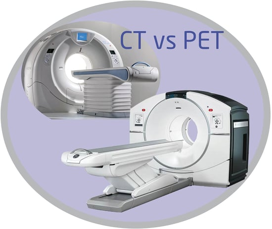 best. Cancer cells grow quickly, taking up larger amounts of sugar or glucose than normal cells. In order to “see” cancer and its growth, a patient will either drink or be injected with a sugary tracer before the PET Scan. The PET Scanner will then trace this form or radioactive sugar as it is absorbed by the body’s cells.
The scanning process is fairly simple: a patient rests on a table and slides into a large scanner that’s tunnel-shaped. It’s an outpatient procedure that is painless. The length of time for the scan varies depending upon which part of the patient’s body is being evaluated.
The Difference Between a PET Scan and a CT Scan
The main difference between PET and CT scans is the technology used to create the images. PET scans use a radioactive tracer to detect metabolic activity, while CT scans use X-rays to create detailed images of the body’s anatomy. During a CT scan, the patient lies on a table that moves through a donut-shaped machine that takes X-ray images from different angles. These images are then combined by a computer to create detailed cross-sectional images of the body. CT scans are particularly useful for detecting abnormalities in the bones and soft tissues, as well as for guiding procedures like biopsies and surgeries.
A combination of PET and CT imaging can reveal information about both the function (PET) and structure (CT) of cells and tissues in the body in a single imaging session. This combination scan enables physicians to examine medical conditions and abnormalities at a cellular level.
During a PET/CT scan, a patient will receive an injection of a radioactive tracer, which is absorbed by specific tissues like areas of inflammation or cancer cells. The tracer emits positrons, which collide with electrons in the body, producing gamma rays that are detected by the PET scanner.
The CT scanner uses X-rays to produce details images of a body’s anatomy. A computer combines the PET and CT images to provide a highly detailed picture of the body’s metabolic and anatomical features.
A PET/CT scan provides several advantages over traditional PET scans or CT scans alone. It provides better accuracy in detecting cancer, better localization of lesions and more precise staging of cancer. It also allows for more efficient diagnosis and treatment planning. However, PET/CT scans do involve exposure to ionizing radiation, which can be a concern for some patients. It’s also possible to have an allergic reaction to the radio-tracer or to experienced minor pain. Some procedures require a catheter to be placed in the patients bladder, which can cause temporary discomfort.
What Is a PET/MRI?
A PET/MRI scan is a hybrid imaging technique that combines the metabolic information from a PET Scan with the detailed anatomical information from an MRI scan. This type of scan provides highly detailed images of the body, allowing physicians to diagnose and monitor a wide range of conditions, including neurological disorders, cancer and heart disease. It shows soft tissue more clearly, so it’s easier to identify soft tissue cancers in the legs, pelvis, arms and upper body.
Talk To An Expert
Are you looking for a new CT Scanner, PET Scanner or MRI Scanner? Talk to the experts at Atlantis Worldwide. For more than 30 years, clients have found ideal medical imaging solutions to fit their needs and budgets by purchasing used or refurbished medical imaging equipment. Find out if this could be the right solution for you.
Contact Us Today!
Some blogs you may have missed:
- CT Scanner Slice: Handy Guide
- Comparing C-Arms: The Ultimate Guide
- Selling Your Used Medical Imaging Systems vs Trading-in
- Extend Your CT Tube Life by 40% or Greater
- CT Scanner: Air Cooled vs Water Cooled
- Free CT Resources
About the author: Vikki Harmonay