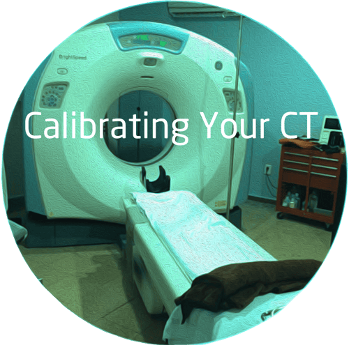
CT scanners are the workhorses of imaging departments around the globe, being used in therapeutic, diagnostic and interventional procedures. In fact, there are more than 75 million CT scans per year in the U.S. The most common CT scans are done on the neck, head and facial regions, although there has been an increase in the number of cardiac, angioplasty and neuro CT studies.
Because there are so many CT scans performed, it is important to calibrate your CT unit on a routine basis. This will guarantee that your scans will have accurate details in the high resolution images created of arteries, nerves and other vascular structures.
What Is CT Calibration?
To calibrate a CT scanner, you need to scan an object or phantom with a known radio density in order to check whether the measurements are giving the appropriate HU or Hounsfield units. While there are no hard and fast rules about how often you need to check your scanner’s calibration, it is important to execute them regularly.
What Is A HU Value?
The HU value measures the absorption/attenuation of radiation within tissue. A grayscale is produced during the CT image reconstruction phase. It reflects the appropriate HU’s for the study that’s done. More than 200 shades of gray can be rendered by a CT scanner, ranging from the lightest whites to the darkest grays. The slightest difference within 2% in tissue density can be detected with this kind of contrast. In order to maintain this level of accuracy without image distortion or loss of value, a regular CT calibration must be performed. Without it, image distortion or lack of proper contrast could lead to a misdiagnosis or delay in treatments for patients that are critically ill.
Calibration is also important if you are testing a CT scanner for purchase. This allows the buyer to test the accuracy of the scanner for studies at their practice.
There are three things that affect the calibration:
- Exposure factors used
- Technological characteristics
- Image viewing conditions
Parameters Tested During A CT Scanner Calibration
- The calibration will measure the homogeneity of an image. It’s essential that hardening artifacts are avoided. Plus, the tissue that’s being imaged should have a uniform appearance that’s free from artifacts
- It will calculate the relevant number of different tissues in a body
- It will measure noise, or the unwanted densities or values in an otherwise homogenous image
- Linearity is measured with a phantom with several materials that mimic different tissues, then those images are compared with the actual HU of the tissue
- Spatial resolution will be measured. A scan must be able to distinguish the differences between two structures with different densities, like fat cells versus muscle tissue
- Low contrast resolution will be tested, so CT imaging can distinguish between more than 200 different shades of gray
- Slice thickness and the positioning of the couch (the movement of the table during the scan will also be tested, in 1mm to 4mm increments
At this time, scientists are still searching for a universal standard for calibrate scanners. Until then, your bio team should work closely with department imaging directors and radiologists to determine the correct protocol during calibration.
Talk To An Expert
It’s always wise to talk to an expert about how often you should calibrate your CT scanner. The team at Atlantis Worldwide is happy to help you with this and any other questions about medical imaging devices, protocols and testing.
Follow Atlantis Worldwide on Twitter: @AtlantisLLC
Other blogs you may have missed:
- Is Your CT Tube About To Fail?
- It's All About CT Detectors
- CT Scanner Artifacts- How Do I Correct Streaks?
- Should your business lease or buy medical imaging equipment?
- Free CT Resources
Meet the author: Vikki Harmonay


