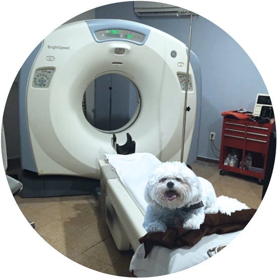
As general practitioners, veterinarians face a lot of challenges. They deal with literally every aspect of veterinary medicine and serve as the front line of care for their patients with fur, fins, scales and feathers. Since they are general practitioners, do they really need to invest in an in-house CT Scanner? The experts at Atlantis Worldwide have prepared this list of questions to help veterinarians decide if a CT Scanner is right for you and your practice.
Will It Be Profitable To Your Business?
CT Scanners are expensive and also involve a big investment in training, maintenance and space. If you choose a fixed CT Scanner, the costs are even more, as the scanning room has to be completely shielded. The big question is, will your clinic be performing enough CT scans to make it profitable for your business?
How Much Do You Know About CT?
Are you familiar with the proper protocol and positioning for CT scanning? Do you understand the correct indications for CT scanning and the limitations of the technology? Do you know how to identify artifacts and troubleshoot a CT during a scan if needed? This knowledge is essential if you’re going to have a CT Scanner in your office.
In addition, the quality of the CT study will greatly influence its diagnostic value. Because your patients must be anesthetized for a CT scan, you need to get the diagnosis right the first time. For example, if performing a thorax scan, it must be done with a breath hold to decrease atelectasis. If there is a pleural effusion, the patient may need a thoracocentesis before the scan if it’s safe to perform one on the pet.
Would You Need A Cone-Beam CT or Slice CT?
Which CT scanner would be best for your practice?
Cone-Beam CT:
A cone-shaped X-ray beam is detected by a flat panel sensor, with both the X-ray beam and sensor rotation around the patient, which creates a larger volume of data in a rotation.
A cone beam creates a single larger volume of data that’s then reconstructed. That means it’s more susceptible to motion artifact than a slice CT. If there happens to be transient motion, it affects all of the images. Because a cone-beam CT rotates more slowly around the patient, it’s inherently more susceptible to motion artifacts.
Cone-beam CT scans a larger section at once, so there’s less collimation and more stray X-ray beams getting registered on the plate. The bigger the body part image, the more scatter. For example, there would be more scatter for a large dog abdomen or thorax when compared to a cat. This increase in scatter leads to decreased contrast resolution, or a decreased ability to distinguish different shades of gray.
A big advantage of the cone-beam CT is the decreased radiation dose to the patient. It also takes up less space, costs less to purchase and offers better spatial resolution when compared to slice CT.
Slice CT Scanner:
In a slice CT Scanner, a narrow beam rotates around the pet and is detected by an array panel sensor. This creates a series of thin slices. Slice CT emits collimated X-rays in thin slices through the pet that are detected by the opposing detector. With slice CT, artifacts appear only in the slices that have motion.
It’s important to note that a slice CT takes up more space that a cone-beam CT and it also delivers a higher radiation dose than a cone-beam CT. A fixed multi-slice CT scanner must be housed in a room that is at least 14 ft. X 20 ft., but this can vary depending on the size of the machine. It also requires a control area outside of the room so the technician can run the scan. The room and the glass window must be shielded with lead that’s formulated by someone trained in radiation shielding or by a physicist.
Some smaller portable multi-slice CT systems require less space and also require no shielding. These portable CT scanners were developed for humans and come with a smaller gantry size, which limits the size of the patient that can be imaged.
Is It Better To Refer Patients To A CT Radiologist?
There are benefits to having your CT scans done in a facility with an on-site radiologist and other specialists. For example, a radiologist can evaluate the images during the scan, and a specialist could perform aspirates or biopsies of abnormal lymph nodes or obtain other tissue samples while the patient is under the same aesthetic event.
It’s common for patients to have procedures immediately after a scan and while under the same anesthesia, like performing rhinoscopy with biopsies for a patient with nasal mass. By doing the CT scan in a clinic with specialists, the pet won’t have to undergo anesthesia as much.
Should You Buy A CT Scanner?
Before you decide if a CT Scanner is right for your veterinary practice, first reach out to a veterinary radiologist or spend time at an imaging facility with a radiologist to become familiar with the protocol, as well as the positioning and indications for CT. Next, talk to an expert like Atlantis Worldwide. We’ve helped veterinarians determine whether a CT scanner makes sense, and if so, which CT scanner would fit their needs best. We’ve been providing refurbished and used medical imaging equipment for healthcare facilities for almost 30 years and would love to help you with your decision.
Some blogs you may have missed:
- 13 Questions to Ask Your Imaging Equipment Service Provider
- The 101 On Veterinary X-Ray Equipment
- C-Arms - The Perfect Tool for Veterinarians
- Does Your Veterinary Practice Need a MRI?
- Choosing A C-Arm For Your Veterinary Practice
Meet the author: Vikki Harmonay


